Canine Parvovirus
- Canine parvovirus (CPV) is a
highly contagious diseases of young dogs characterized by acute
hemorrhagic enteritis, leukopenia and occasionally myocarditis
ETIOLOGY
- Canine parvovirus – SS DNA
- Several antigenically different
serotypes:
- CPV-1: Minute virus of canines
– causes myocarditis and respiratory diseases and intestinal enterocyte
hyperplasia with eosinophilic to amphophilic INIB
- CPV-2: Subtypes CPV-2a, and
CPV-2b – causes parvovirus enteritis
- Genus: Protoparvovirus
- Family: Parvoviridae
- Parvo: from Latin, ‘small’
Host
• Young (less than 6 month),
unvaccinated or incompletely vaccinated dogs are most susceptible.
• Cat, mink, calves can also affected
Transmission
• Direct contact
•
Virus
shed in feces of infected dogs
•
Contaminated
food, water, fomites
Pathogenesis
• Virus reach to nasopharyngeal mucosa→
viral uptake by epithelium over tonsils and Peyer's patches → Virus (1-2 days)
→ Lymphoblast dissemination to many tissues (3-4 days) → Lymphocytolysis
(Leukopenia) → releases virus, causing viremia → neutralizing antibody -
terminating viremia (5-7 days) →Infection of GI crypt epithelium and Peyer's
patches (5-9 days)
• Further diseases development depends
on amount of virus , lymphocyte
lysis and the rate of cell proliferation
in the crypts of Lieberkuhn (Lieberkuhn is the pit between
the villi in the small intestine)
• Virus reach to crypts of Lieberkuhn→ Replication of virus in actively dividing enterocyte ( Enterocyte : intestinal absorptive cells, are simple columnar epithelial cells which line the inner surface of the small and large intestines.) → Lysis, necrosis and sloughing of enterocyte → Villus atrophy, erosion or ulceration → Hemorrhages and malabsorption → bloody diarrhea → Dehydration
• Epithelium regeneration → Facilitate
virus replication → more destruction If stem cells present -
Regeneration occurs
• Cytolysis of proliferating cells in
the bone marrow causes myeloid and erythroid hypoplasia (Megakaryocytes are the
least sensitive to lysis)
• In new born puppies → Cardiac
myocytes are actively dividing cells up
15 day age → Replication of virus in actively dividing cardiac myocytes → Lysis
and necrosis of cardiac myocytes →
Myocardial necrosis → arrhythmia and unexpected Death
Clinical
Signs
•
Three forms of disease:
❶ Generalized:
•
Pups less than 2 weeks
- Found Dead – Rare
❷Cardiac form:
•
Pups 2-8 weeks
- sudden death from cardiac arrhythmias
•
Survivors usually die by 5 months of age from chronic
myocardial fibrosis and resultant cardiac arrhythmias.
❸Leukopenia/enteritis:
•
Common form in pups 8 weeks and older
•
Vomiting, anorexia, pyrexia, dehydration, lethargy,
bloody diarrhea.
•
Leukopenia, with relative or absolute lymphopenia,
hypoproteinemia, and anemia.
•
Myocardial and enteric forms rarely occur together.
Macroscopic Pathology
Leukopenic/enteric
form :
•
Segmental to
diffuse hemorrhagic gastroenteritis with mucoid/bloody intestinal contents and
fibrinous serosal exudate
•
Peyer's
patch necrosis
•
Enlarged,
congested, edematous mesenteric lymph nodes
•
Semiliquid,
yellow-gray bone marrow
•
Thymic
atrophy
Myocardial
form:
•
Pale streaks
in myocardium, pale, flabby
•
Pulmonary
edema with white frothy fluid in trachea and bronchi
 |
Canine small intestine dilatation, luminal hemorrhage
 |
Segments of the small intestine are diffusely reddened (active hyperemia of the mucosa), and the serosal surface is roughened, faintly granular, and petechiated.
 |
Note the multifocal pale areas (arrow) in the ventricular myocardium
Microscopic Pathology
• Severe necro-hemorrhagic gastroenteritis; crypt necrosis; blunting; fusion
• Basophilic intranuclear inclusion bodies in GI epithelium are variably evident.
• Necrosis and depletion of lymphoid tissues
• Bone marrow depletion
• Focal myocardial necrosis with intranuclear basophilic inclusion bodies in cardiomyocytes
 |
Parvovirus infection, section of myocardium. An intranuclear basophilic inclusion body is in a myocyte (arrow)
Diagnosis
•
Clinical
signs
•
Histopathology
•
Laboratory tests
•
Virus
neutralization tests
•
Enzyme-linked
immunosorbent assay (ELISA)
•
Polymerase
chain reaction
•
Serological
tests
Canine Parvovirus Antigen Test Kit
Sample Information
● Samples must be at room temperature (18-25°C) before beginning the test procedure.
● Canine fecal matter is required for this test. Swabs are provided for sampling.
● Fecal samples can be stored at 2-8°C for 24 hours. If longer storage is required, samples should be frozen.
Test procedure
1. If stored in a refrigerator, allow all components to equilibrate at room temperature (18°C-25°C) for 30 minutes. Do not heat.
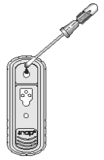
2. Obtain a sampling swab and a SNAP device for each sample to be tested. Pull and twist the tube covering the swab tip to remove the tube from the swab/reagent bulb assembly (A). Using the swab, coat the swab tip with fecal material. Then, return the swab to the tube (B).
3. Break the purple valve stem inside the bulb assembly by bending the assembly at the narrow neck (C), rebending the opposite way may be helpful. Squeeze the reagent bulb three times to pass the blue solution through the swab tip and mix it with the sample (D).
4. Place the SNAP device on a horizontal surface. Using the swab as a pipette, dispense 5 drops of the fluid into the sample well, being careful not to splash contents outside of the sample well.
The sample will flow across the result window, reaching the activation circle in 30-60 seconds. Some sample may remain in the sample well.
5. When color FIRST appears in the activation circle, push the activator firmly until it is flush with the device body.

NOTE: Some samples may not flow to the activation circle within 60 seconds, and, therefore, the circle may not turn color. In this case, press the activator after the sample has flowed across the result window.
6. Read the test result at 8 minutes.
Interpreting Test Results
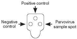
To determine the test result, read the reaction spots in the result window and compare the color intensity of the sample spot to that of the negative control spot.

Positive Results
Color development in the sample spots that is darker than the negative control indicates a positive result, and the presence of parvovirus antigen in the sample.

Negative Results
Color development only in the positive control spot indicates a negative result.

TREATMENT
- Intravenous
fluids (a drip) to treat shock and correct dehydration and electrolyte
abnormalities
- Anti-sickness medication
- Plasma
transfusions and/or blood transfusions to replace proteins and cells
- Antibiotics
to treat or prevent secondary infections as a result of the effects of parvovirus
infection like The most
common antibiotics used include ampicillin, cephalexins, and flouroquinolones
- Tube
feeding


Very Nice &Very Helpful👌👌
ReplyDeleteVery helpful information...Thank you🤝🤝
ReplyDeleteThank you and Welcome Dr
DeleteVery nice Dr
ReplyDeletePost a Comment