Introduction
·
Bovine Tuberculosis is a disease caused by bacteria Mycobacterium bovis.
· TB usually affects cattle and all mammals causing a general
state of illness, coughing and eventual death.
· Tuberculosis can be transmitted from animals to humans as
well as to other animals also so it is zoonotic disease.
·
The bacteria M. tuberculosis, that is different
the type that usually causes disease in humans.
·
While M. bovis is a
different type of bacteria to causes disease in animals.
· The name Tuberculosis(TB) comes from the nodules, called “
tubercles ” which form in the lymph nodes of affected animals.
·
They
are generally killed at a temperature of 60oC for 15 minutes
· The bacteria of M. bovis survive in a wide range of alkalis
and acids. And also long periods in moist and warm soil.
·
M. bovis in faeces and they survive 1 – 8 weeks.
·
The bacteria of M. bovis is killed in sunlight.
·
Bovine tuberculosis is a zoonotic disease and causes tuberculosis in
human.
ETIOLOGY
·
Etiology: Mycobacterium bovis.
·
Human Tuberculosis : M. tuberculosis, M. africanum (Africa), M. canettii
·
TB in goats : M. caprae
·
TB in avian : Mycobacterium avium
·
TB
in small rodents : M. microti
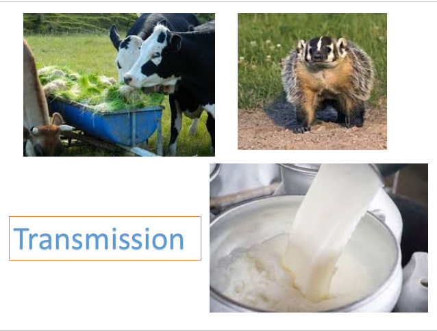
TRANSMISSION
·
Infected
domestic animals like cattle, wild animals and badgers
·
Breath
·
Raw Milk (Raw milk, but pasteurisation milk prevents the spread.)
·
Discharging lesions like saliva, urine.
·
Contamination
of fodder
·
Respiratory droplets or within dust particles that can travel long distances
in the air in TB.
·
Spread by direct wound during slaughter and hunting of animal.
·
Vertical transmission can also reported in infected females.
HOST
·
Mycobacterium
tuberculosis (TB) has been reported in All domesticated and non-domesticated
animals
·
Also Wild mammal species including lions, tigers,
leopards.
·
High infection rate in badgers.
·
Badgers are a significant source of Tuberculosis in
cattle.
Which animals are more susceptible for TB ?
·
poorly nourished Animals
·
Stressful is also susceptible for TB
·
Younger cows and growing heifers are most at risk.
·
Evidence more in dairy farms animals have a higher
risk of infection.
·
In cattle, excretion of M. bovis begins
around 87 days after infection occurs.
Pathogenesis
·
Cattle,sheep
and goats are infected through inhalation of fomites and fluid droplets
contaminated with the bacteria or ingestion
via the milk or colostrum
·
These droplets
are deposited in mucus layer of the respiratory system and are subsequently
phagocytosis by alveolar and tissue macrophages
·
These
Macrophages appear to use several routes to spread the M.Bovis across mucus layer and then to regional lymph nodes and the
lung.
·
In tonsillar
mucosa the macrophages are cross the mucosal barrier and migrateor bind
with the tonsil, and spread the bacteria
to tonsillar tissues.
·
In pharynx
mucosae the macrophages encounter and phagocytose bacteria à spread them to local lymphoid tissues through
afferent lymphatic vessels to regional lymph nodes such as the retropharyngeal
and parotid nodes
·
Finally the
bacteria reach in bronchi and bronchioles and bacteria are deposited in the
mucus layer and phagocytosed by alveolar macrophages and spread to local
lymphoid tissues (BALT) through afferent lymphatic vessels to regional lymph
nodes like the tracheobronchial and mediastinal nodes and infection occurs
·
spread the
infection of M.Bovis other regional
or system like liver, spleen, lymph nodes, and intestines, by the leukocyte in
the blood or lymphatic vascular systems.
·
Bovine TB may be sub acute or chronic, with a variable rate of
progression.
·
A small number of animals may become severely affected within a few
months of infection and while some animals may take several years to develop
clinical signs.
·
The bacteria can also lie in the host without causing disease for a long
periods.
·
Cattle
usually show no clinical signs to TB unless the disease has affected multiple
organ systems and is very advanced which is quite rare.
·
Typically,
infected cattle are asymptomatic and are only detected by skin testing or at during
slaughter.
The usual clinical signs include:
·
Weakness,
·
Loss of appetite and weight,
·
Fluctuating fever,
·
Dyspnoea,
·
Intermittent hacking cough,
·
Signs of low-grade pneumonia,
·
Diarrhoea,
·
Enlarged and prominent lymph nodes.
·
Tubercular mastitis is one of the common
conditions in M. bovis infection.
Macroscopic Pathology
·
Characteristic gross lesion of
an animal infected with bovine TB is the presence of “tubercles” within the
body
·
A tubercle is a white nodule
usually 1mm-2cm in diameter within a lymph node or organ. Commonly found in the
thoracic cavity
·
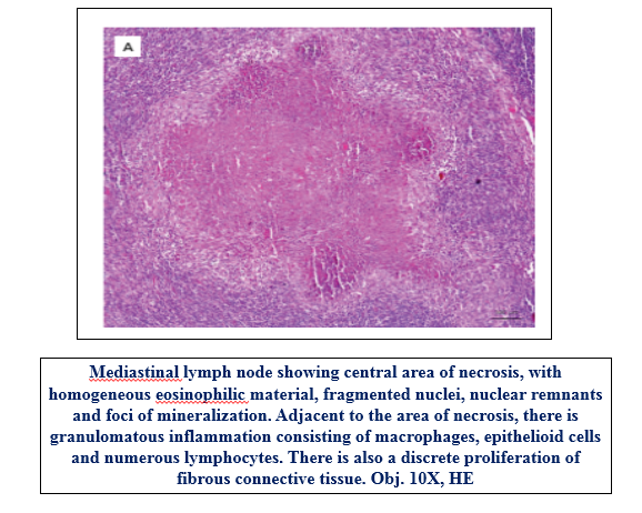
The centre of the granuloma is
usually necrotic and calcified.
·
Moderate splenomegaly seen.
·
Chronic pneumonia with or without regional lymph node involvement.
·
Caseous necrosis is prominent in the granulomas of bovine tuberculosis
MICROSCOPIC Pathology
·
Granulomatous inflammation is the hallmark of infection with
·
Lymphocytosis and monocytosis observed in case of TB
Diagnosis
·
Base on clinical sign and macroscopic and microscopic lesions
·
Sample collection:
Sputum,
milk, uterine discharges, pleural and peritoneal fluids, urine, faeces and
Tissue.
·
Microscopical examination
o
Z-N
stain(Ziehl-Neelsen method)- The
tubercle bacilli appear in clumps as slender rods stained pink in a blue
background.
o
In milk
samples, the organisms are commonly seen in epitheloid cells.
·
Cultural examination: Culture isolation of
the causative agent from fresh tissue or other specimens
M.
bovis isolates - Stone
brink's medium
·
Animal inoculation
M.
bovis- Guinea pigs, Rabbits
M.
tuberculosis- Guinea pigs, Rabbits
M.avium-
Chicken,Rabbit
·
Tuberculin
Test (Delay type Hypersensitivity test)
o
In live cattle, pig, deer tuberculosis is
usually diagnosed in the field with the tuberculin skin test.
o
Tuberculin / purified protein derivative (PPD)-
is a extracts of M. bovis that is used in skin testing in animals and
humans to identify a tuberculosis infection.
o
Purified protein derivative (PPD) is complex
mixture of protein, lipid, carbohydrate, nucleic acid
o
Intradermal test:
The side of neck shaved à Skin
fold Measure by vernier caliper à0.1 ml of tuberculin (PPD) is injected i/d at
sides of neck. (caudal fold of tail, vulvar lip) à After
72 hours edematous swelling
àindicates
positive reaction.
àThickness
of swelling 3mm is doubtful and 4mm is positive.
·
I/D comparative cervical: The side of neck shaved
o
Skin fold Measure à0.1
ml of tuberculin (PPD) of bovine and 0.1 ml tuberculin of avian is injected i/d
into different side of neck.(12 cm apart)
o
After 72 hours edematous swelling indicates positive reaction.
o
Thickness of swelling 3mm is doubtful and 4mm is
positive at the site of bovine PPD injected
o
False
positive reaction- Tuberculin test may be attributed to
sensitization to mycobacteria other then M. bovis. So comparative i/d test
use in preference to single i/d.
o
False
Negative – Test perform before 30 days post infection.
o
Some cattle unresponsive referred to as anergy
o
Immunosuppression may contribute to the inability to respond to the tuberculin test
·
Ophthalmic: One
drop of tuberculin is dropped into conjuctival sac and again 3 drop after 48
hr. After 24 hour of second instillation, the eye should show purulent
conjunctivitis in a positive.
·
PCR
·
ELISA
TREATMENT
•
On December 28th, 1908 the French bacteriologists
Albert C. Calmette and Guérin notified a loss of virulence of M. bovis
when cultured in bile containing media.(Bacille Calmette-Guerin)
•
A live attenuated vaccine (BCG)
is the current vaccine for tuberculosis
•
BCG name due to Bacille
Calmette-Guerin
•
Isoniazid, Rifampicin, Pyyrazinamide
& Ethambutol drugs are used.
References:
Pathologic Basis of Veterinary Disease Expert Consult
6th edition


Very nice information.
ReplyDeleteThank you Dr
DeleteBeautiful yaar
ReplyDeleteThank u so much Dr
DeleteVery nice information dr.... Keep it Dr 👍
ReplyDeleteThank you so much Dr
DeletePost a Comment