Rabies
- Rabies is a neurological, invariably fatal, viral disease of mammals characterized by nervous signs, salivation, paralysis , non suppurative encephalitis and intracytoplasmic inclusion bodies (Negri bodies)
- Many
islands countries are free from rabies like United Kingdom, Ireland,
Sweden, Norway, Iceland, Japan, Australia, New Zealand, Singapore, most of
Malaysia, Papua New Guinea, the Pacific Islands etc…
- Synonym :
- Hydrophobia ( Animal which
fear by water in case of rabies )
- Lyssa ( Due to caused by Lyssa virus )
Etiology
• Rabies virus (Genotype
1) – Neurotropic
• Genus: Lyssavirus
• Family: Rhabdoviridae
• Total 11 distinct species can be distinguished within the genus in
lyssavirus
Two
biotypes
❶ Fixed virus: ❷
Street virus:
•
Laboratory
vaccine virus Produced natural occurring diseases
•
Tropism to CNS Tropism
to CNS and salivary glands
•
Not secreted in saliva Secreted in saliva
•
No Negri bodies
Negri bodies
in 70 % cases
• Marked neuronal denegation
• Adapted to CNS by passage
Host
• All warm blooded animals /mammals
(wild and domestic)
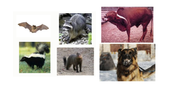
• Jackals in India are reservoir
vectors
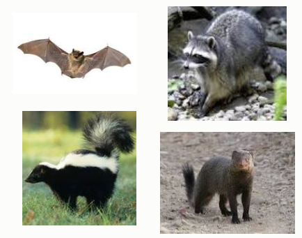
• Bats, foxes, skunks, raccoon,
mongoose are reservoir vectors in other part of world
Transmission
• Direct contact
Saliva of infected animals
• Rarely reported other route of
transmission of rabies like….
•
Aerosol
route of transmission in laboratories
and bat caves
•
Sometimes
Organ transplantation like cornea, pancreas, kidneys and liver
•
Also
seen Transmission in a human infant and milk to a lamb
•
Ingestion
of infected animals so Not recommended to consume tissues from rabid animals.
(CFSPH,2009)
Pathogenesis
Virus enter
in to body via bite of infected animals
•
Initial
multiplication in muscles (Which can enter peripheral nerves directly –
Short incubation period ) à And enters into peripheral nerves à Ascends (retrograde axonal
transport the peripheral nerve (12 to 100 mm/day) à Enter to the dorsal root ganglion à Enters the spinal cord à Ascends to the brain via ascending
and descending nerve fiber tractsà reach to Infects brain cellsà Spreads to salivary glands and the
eye, replication and is excreted in saliva à
•
Viral
envelop G protein → bind to muscles →
virus replication muscles → virus bud out from muscles → bind to acetylcholine receptors at the neuromuscular junction →
pinocytosis (it is a process by which
liquid droplets are ingested by living cells and it is one type of endocytosis
and engulf external substances, gathering them into special membrane-bound
vesicles contained within the cell.) → virus in vesicle enter in to nerve and Virus
induced neuronal apoptosis and degeneration.
Incubation Period
• It is depends on
•
Amount
of virus transmitted
•
Virus
strain
•
Site
of inoculation (bites closer to the head have a shorter incubation period)
•
Host
immunity
•
Nature
of the wound
• In Dogs and cats - 10 days to 6 months
•
Most
cases - 2 weeks and 3 months
• In Cattle - 25 days to more than 5 months
• In Humans - few days to several
years
•
Most
cases become apparent after 1 to 3
months
Clinical Signs
• Signs in domestic animals are
similar with some differences between species
•
Prodromal
phases
•
Excitatory
phase (furious form)
•
Paralytic
phase (dumb form)
❶Prodromal phases:
• Animal can have a subtle change in
temperament ( Change the behavior )
• Restlessness, anorexia or an
increased appetite, vomiting, a slight fever.
• Dilation of the pupils
•
Hyper
reactivity to stimuli and excessive salivation
❷Excitatory phase (furious form):
•
Infection
of limbic system of brain mainly Hippocampus
and other
•
In
furious form - Excitatory phase
predominant
•
Cattle,
Carnivores, Sheep, Pig, Cat seen the furious form
•
Wandering,
crying, polypnea, drooling and attacks on animals, people or inanimate objects
•
Often
swallow foreign objects such as sticks, stones, straw or feces
•
Cattle with
rabies have unique bellowing, general straining, tenesmus( Rectal straining ) and signs
of sexual excitement
❸Paralytic phase (Dumb form):
•
It
is associated with infection to
cerebellum
•
Excitatory
phase is extremely short or absent
and the disease progresses quickly to the paralytic phase
•
Throat
and masseter muscles become paralyzed so unable to swallow
•
Facial
paralysis / lower jaw may drop due to paralysis
•
Ascending
spinal paralysis (starting from tail)
•
Death
usually occurs within 2 to 6 days, as the result of respiratory failure
•
In
human seen the Hydrophobia
Macroscopic
Pathology
•
There
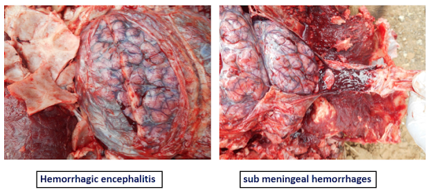
are no gross lesions
•
Cattle
and horses may see hemorrhage in the spinal cord
•
Evidence
of injury or self-mutilation
•
Presence
of unusually material or foreign bodies in stomach/alimentary tract.
Microscopic
Pathology
•
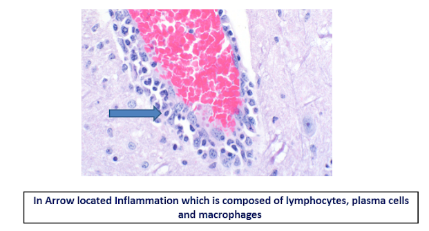
Nonsuppurative encephalomyelitis, with ganglioneuritis and parotid
adenitis
•
Perivascular cuffing, Babe’s nodules and Negri bodies are pathognomonic
for rabies
•
Lesions
most severe from the pons to the hypothalamus, and the cervical spinal cord;
medulla generally not affected
•
Perivascular
lymphocytic cuffing; perivascular hemorrhage; leptomeningitis
•
Babe’s (Glial) nodules – composed of Microglial cells
•
Neuronal
degeneration and inflammation is most severe in carnivores and mild in pigs and
herbivores
•
Negri
bodies are round to oval, 2-8 um,
eosinophilic intracytoplasmic inclusions
•
Aggregations
of strands of viral nucleocapsid; transforms into a granular matrix
•
Negri bodies - hallmark of rabies infection
•
Carnivores
– hippocampal neuron
•
Herbivores - Purkinje neuron of cerebellum
•
Negri
bodies are rarely found in the ganglion cells of the adrenal medulla, salivary
gland and retina.
•
Early
infections and up to 30% of rabies infections Negri bodies not found
•
A
spongiform lesion with vacuolation of gray matter neuropil, similar to scrapie,
was originally described in foxes and skunks with rare reports in the horse,
cow, cat and sheep.
•
Parotid
adenitis
•
Epithelial
cell degeneration
•
Lymphoplasmacytic
inflammation
Diagnosis
•
Base on Clinical signs.
•
Laboratory
tests like…
•
Fluorescent antibody test (FAT)- recommended by
both WHO and OIE - ‘Gold-standard’ test
•
Immunoperoxidase methods
•
Enzyme-linked immunosorbent assay (ELISA)
•
Mouse inoculation test
•
RT – PCR
Treatment
•
There is
no treatment of Rabies
•
We prevent
the Rabies by Rabies vaccine.
•
Sometime Rabies
vaccine is also failure due to not maintain cold chain and faulty vaccination.
•
Post bite
rabies schedule is…
1st vaccine dose 0 day (At time
of bite )
2nd vaccine dose 3 day
3rd vaccine dose 7 day
4th vaccine dose 14 day
5th vaccine dose 28 day and
Some veterinarian given on 90 day also.
But in field condition we given up to 28
day.
Rabies Virus Ag Test
·
Rabies Virus Ag Test is a sandwich lateral flow
immunochromatographic assay for the qualitative detection of rabies virus
(Rabies Ag) in animal’s saliva or brain tissue.
Time: 5-10 min
Sample for test:
Saliva and Brain sample
PRINCIPLE: Quicking Rabies Virus Ag Test is based on sandwich lateral
flow immunochromatographic assay. The test device has a testing window. The
testing window has an invisible T (test) zone and C (control) zone. When sample
is applied into the sample hole on the device, the liquid will laterally flow
on the surface of the test strip. If there is enough Rabies virus antigen in
the sample, a visible T band will appear. The C band should always appear after
a sample is applied, indicating a valid result. By this means, the device can
accurately indicate the presence of Rabies virus antigen in the sample.
PROCEDURE
1. Collect
animal’s saliva or 10% brain tissue fluid homogenates with the swab stick.
2. To
collect an animal’s saliva, gently rub the cotton swab inside the mouth, along
the gums and cheeks, making sure the swab is sufficiently wet.
3. Unscrew
the cap from the provided assay buffer tube and insert the wet swab.
4. Agitate
it to ensure good sample extraction.
5. Take
out the cassette from the foil pouch and place it horizontally.
6. Use
the pipette to extract solution from the assay tube and transfer 3 drops of
sample extraction into the elliptical sample hole on the test cassette.
7. Interpret
the result in 5 - 10 minutes. Result after 10 minutes may be considered invalid.
RESULTS
Positive: The presence of both C band and T band, no matter how faint the T band appears.
Negative: Only clear C band appears
.
Invalid: No
colored band appears in C zone, no matter whether T band appears.


Usefull information
ReplyDeleteHussain khan best notes on rabies read till yet 👌👌
DeleteFull of knowledge...included every point...
ReplyDeleteThanks Dr. I hope u got everything in this topic.
DeleteUse ful information.. fabulous job...
ReplyDeleteThank you so much Dr
DeleteAmazing content....very useful...Thank You so much👌🤝🤝
ReplyDeleteNeed Rinderpest disease info!!
ReplyDeleteUpdates soon
DeleteVery good information
ReplyDeleteNeed metabolic diseases..Milk fever,post parturient haemoglobin urea,grass tetany
ReplyDeleteUpdated soon...
DeletePost a Comment