Clostridium diseases in animals
1. Black quarter :- Click here
2. Tetanus:- Click here
3. Enterotoxaemia :- Click here
4. Bacillary haemoglobinuria:- Click here
5. Botulism
6. Braxy
7. Infectious Necrotic hepatitis
8. Malignant oedema
|
Organism |
Disease |
|
Cl. chauvoei |
Black quarter/ black leg |
|
Cl. tetani |
Tetanus |
|
Cl. perfringens |
Enterotoxaemia |
|
Cl. septicum |
Braxy, malignant edema (gas agangrene) |
|
Cl. haemolyticum |
Bovine bacillary haemoglobinuria / red water disease |
|
Cl. novyi |
Black disease |
|
Cl. botulinum |
Botulism |
BLACK QUARTER
SYNONYMS:
ü Black leg
ü Quarter ill
ü Symptomatic
anthrax
ü Emphysematous gangrene
ü Charbon symptomatique
ü Felon
ü Carbon oil
symptomatico
ü Raush brand
INTRODUCTION
⇒ Black quarter is caused by clostridium chauvoei and This is an acute infection but not contagious disease of cattle, sheep and goat. The disease is characterized by development of focal gangrenous and emphysematous myositis. This give rise to crepitation and sero hemorrhagic swelling in the heavy muscles like gluteal muscles.The disease produces severe toxaemia with very rapid course and high mortality.
DISTRIBUTION
⇒ Widespread in tropical countries mainly.
⇒ The disease spread rapidly following heavy rainfall.
⇒ In India the disease is sporadic in nature. The disease appears in almost all states of the country during rainy season and warm month (spring to autumn)
ETIOLOGY
⇒ Black quarter is caused by clostridium chauvoei , gram positive rod shaped , spore forming toxin producing anaerobic bacteria.
⇒ The spores are very much unaffected to hot drying and disinfectants and spores can with stand 120 degrees Celsius temperature for 10 minutes.
⇒ The spores can persist in the soil for number of years.
⇒ False Black quarter can be caused by Cl. Septicum and Cl. Novyi.
⇒ Cl. Chauvoei is considered as the primary cause of black quarters and other anaerobes are supposed to be secondary invaders.
⇒ In pig: gas gangrene is due to Cl. Septicum & clostridium chauvoei.
SUSCEPTIBLE HOST
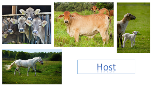
⇒ Cattle is the most susceptible host but the infection may spread to other animal do too traumatization of the muscles.
⇒ The disease may occur in sheep Buffalo and goat.
⇒ Cattle of all breeds are susceptible but the incidence is more common In cattle having 4 to 24 months of age group and good body condition.
MODE OF TRANSMISSION
⇒ The disease spreads from contaminated soils. The contamination of soil is due to infected carcasses which cause pollution of the land. The organisms gain entry through ingestion of infected feeds or possibly through contamination of wounds. Organisms are excreted in the faeces.
⇒ Most of infection transmitted by following skin wound
·
Wound during shearing
·
Wound during docking
·
Wound during vulvar/vaginal
laceration at time of parturition or lambing
PATHOGENESIS
Ingested
organisms are carried from the intestine via circulation to the skeletal
muscles.
⇓
The
spores from the elementary tract penetrate tissues from the places of breach of
alimentary mucosa due to trauma.
⇓
Some
of the sports in the muscles are destroyed by phagocytosis and others remain latent for at least several weeks.
⇓
The
infection may affect the muscles inter muscular tissue.
⇓
Very
often heavy muscles which are well formed like muscles of gluteal region, loin and shoulders are affected.
⇓
There
is necrosis of the muscles and blood capillaries.
⇓
Gases
used to accumulate within the muscle fibres due to fermentation.
⇓
The
infection can also be spread through peritoneum and pleura.
⇓
Exotoxins
produced from the organisms which causes systemic reaction characterized by toxaemia and local reaction characterized
by necrotising myositis.
CLINICAL FINDINGS
v Incubation
period is usually 2-5 days. Under the influence of toxic products elaborated
during the growth of the organisms the muscles degenerate and gases are
evolved. The toxic products are absorbed by the body fluids causing systemic
disturbances and a decrease in the animals vitality.
v In cattle:
ü The
first symptom is a rise in body temperature which may be as high as 106 degrees Celsius or 108 degrees Celsius
but sometimes there is hardly any sign of fever.
ü The
appetite is lossed and rumination is suspended
ü There
is stiffness or lameness in one of the Limb. This being the usual early
symptom.
ü Very
soon characteristic swelling develops in one of the thick layers of muscle.
ü Most
commonly the lesions are located on the Thigh,
buttocks, shoulder next and lumbar region and more rarely in the intermandibular space or in the tongue.
ü The
structures get disrupted due to gas pocket giving rise to a spongy texture and
on pressure swellings emit crackling or crepitation sound due to emphysema.
ü There
is laboured breathing and accelerated pulse
rate 100 to 120 per minute.
ü Finally
the temperature drops and the patient dies within 12 to 48 hours after
manifestation of the clinical signs.
v Sheep:
ü Stiff
gait
ü Bleeding
from nose
ü Anorexia
ü Fever
ü depression
ü Severe
lameness in one or more limbs
ü Not
seen sub cutaneous edema and not felt gaseous crepitation sound before death
ü Death
v Horse:
ü Not
well define the clinical signs
ü Pectoral
edema
ü Stiff
gait
ü Incoordination
of gait observed
Macroscopic Pathology
ü Quick
putrefaction and bloat
ü Blood
stained froth seen in nostril and anus
ü Rapid
clotting of blood
ü When
incision at affected muscles mass is dark red to black colour
ü Affected
tissue are swollen, rancid odour and containing gas bubbles
ü Lungs
are congested and atelectasis
ü Lesions are limited to
affected muscles. Muscles of shoulders thigh, Neck are usually affected. Lesions
me also be observed in the tongue diaphragm and myocardium. Large crepitating
swellings are the most characteristic necropsy finding.
Microscopic Pathology
ü Edema, emphysema,
myonecrosis and neutrophilic cellulitis.
DIFFERENTIAL DIAGNOSIS
ü Malignant edema
ü Anthrax
ü Lightning strike
ü Bacillary hemoglobinuria
DIAGNOSIS
ü In the field outbreak, a
tentative diagnosis is made from the history
ü clinical observation
ü Post mortem findings.
LAB
TESTS:
ü Microscopic
examination of smear
ü Cultural
test
ü Biological
test
ü FAT
TREATMENT
ü Penicillin
is extensively used and considered as drug of choice. The antibiotic may be
injected into the affected muscles.
ü Penicillin G
sodium/potassium (44,000 IU/kg IV q6–8h)
ü Clostridium chauvoei antitoxin
ü Surgical debridement of affected
muscles
ü IMMUNIZATION
ü Vaccine:
polyvalent, BAIF


Post a Comment